Skip to content

Lublin Digital Union

You are in the right place.

Using technology, we create together for people.

Lublin Digital Union
supports generations.

Using technology, we create together for people.

For Parents

Sustainable development
is caring for the youngest.

Find out how your child can benefit. Learn how to complete the application form and stay in touch with us.

For Researchers

Lublin Digital Union
is the world of science.

The project assumes an innovative, groundbreaking approach to research by using neural networks to assess the progression of a chronic, progressive and incurable rare eye disease, retinitis pigmentosa.

For Youth

LDU is for you.
For many reasons.

You’ll see more! Your exams matter. Get your eyesight checked. Don’t be indifferent. See how you can help others too!

For Students

Check your eyesight.
For yourself. For others.

Using technology, we will create a diagnostic tool together.

Collaboration that brings results.

University of Life Sciences
in Lublin

University of Life Sciences in Lublin plays a key role in the project, one of whose objectives is to conduct genetic research and develop a model for monitoring disease progression in the context of rare diseases in ophthalmology. The research team from the Department of Biochemistry and Toxicology, led by Prof. Dr. habil. Katarzyna Ognik, is the central unit responsible for carrying out this task.

Particularly significant is the university’s impact, which encompasses a wide range of activities related to genetic research. The task assigned to the University of Life Sciences in Lublin constitutes an integral part of the entire project. Its implementation will be based on advanced digital solutions, contributing to a better understanding and monitoring of the progression of rare diseases in the field of ophthalmology. Through active collaboration with the research team from the Department of Biochemistry and Toxicology, the University of Life Sciences in Lublin plays a crucial role in achieving the project’s objectives and expanding knowledge in the field of medicine.

Read more

Medical University
in Lublin

Medical University of Lublin stands out as a leader in implementing change across multiple levels—local, national, and international. Its role in conducting scientific research is particularly significant, and the use of information technologies and artificial intelligence places it among the world’s leading educational institutions.

The institution is widely recognized both domestically and internationally, confirming its substantial contribution to the development of medical science. By specializing in modern technologies and artificial intelligence, the Medical University of Lublin not only develops innovative models of scientific research but also actively implements them.

Research conducted at the Medical University of Lublin focuses on the field of medicine, where an innovative approach to challenges is supported by advanced technologies. The use of artificial intelligence enables efficient data analysis, which in turn contributes to the development of new diagnostic, therapeutic, and preventive methods.

However, the role of the Medical University of Lublin extends beyond scientific research alone. It is an institution that actively engages in serving society—working to improve public health, promoting medical knowledge, and collaborating with local communities. The university not only educates future specialists but also serves as an important link in building a healthier and better-informed society.

Read more

Lublin University
of Technology

Lublin University of Technology is a leading technical institution, serving as a key academic center in the Central-Eastern Macroregion of Poland.

The Lublin University of Technology actively develops interdisciplinary collaboration, integrating medicine and engineering sciences. Since 2010, it has offered a Biomedical Engineering program at both first- and second-cycle (Bachelor’s and Master’s) levels. Education in this field covers areas such as medical informatics, medical electronics, engineering biomechanics, and biomaterials engineering. Students acquire skills in using modern equipment and diagnostic systems based on advanced technologies, including teleinformatics, electronics, and materials science. Moreover, the curriculum prepares graduates for effective cooperation with medical doctors, encompassing the operation, use, and maintenance of medical equipment as well as diagnostic and therapeutic systems. In this way, the Lublin University of Technology stands out as an institution that not only generates new scientific solutions but also actively translates them into practice, integrating engineering and medical disciplines.

The university enjoys strong recognition among Polish educational institutions, ranking among the national leaders in innovation. This is confirmed by the latest Perspektywy Higher Education Ranking 2022, which awarded it third place in Poland for inventiveness.

Read more

The use of digital solutions and artificial intelligence in medicine – a research project

FAQ - Frequently Asked Questions​

Such studies have not been conducted anywhere else yet. The innovativeness of the project lies in the inclusion of both functional assessments (visual acuity, visual field) and imaging studies of the retinal structure (wide-field fundus photography, OCT and angio-OCT), as well as electrophysiological examinations to evaluate the rate of RP progression. These studies will be unique on a European scale, making it possible to identify potential biomarkers of RP progression over time. In the future, this may prove important for determining the effectiveness of new potential therapeutic methods (e.g., gene therapies).


Myopia is a vital public health problem that affects a significant number of people and can lead to vision loss. It develops slowly and insidiously, with significant progression. It often begins at a young age (from school age up to 25 years of age), disrupting the educational and cognitive processes of children and adolescents and, consequently, their normal development. The treatment of myopia places a substantial burden on the healthcare system in Poland. Refractive errors are among the most common visual disorders worldwide (42%), surpassing eye diseases such as cataract, glaucoma, or retinal diseases.

Myopia is a refractive error in which the image is formed in front of the retina. This condition requires appropriate correction (glasses, contact lenses, surgical procedures). If left untreated, it increases the risk of developing glaucoma or retinal detachment. The number of people suffering from myopia worldwide is estimated at over 2.5 billion. It is predicted that by 2050 the number of people with myopia will reach 5 billion, which will constitute half of the global population. Over the past 50 years, the prevalence of myopia has doubled in Europe and the United States. Currently, as many as 30% of Europeans have been diagnosed with myopia.

In Poland, according to a TNS study conducted in 2012 among a group of 1,000 Polish residents over 15 years of age, it was found that 37% of respondents have vision problems, most caused by myopia (46%)—source: Stankiewicz A., Misiuk-Hojło M., Ulińska M., Sobierajski T.: Stopping the Epidemic of Blindness. The Medical, Social and Economic Picture of Retinal Diseases, DMA Association for Age-Related Macular Degeneration, Polish Association of the Blind, Retina AMD Poland, Warsaw 2012.

However, the most alarming data comes from East Asia, where myopia has become a true epidemic. For the sake of comparison, 60 years ago 10–20% of the Chinese population suffered from myopia—today this figure reaches 90% of adolescents and young adults.

The innovativeness of the project lies in the inclusion of both functional tests (visual acuity, visual field) and imaging studies of retinal structure (wide-field fundus photography, OCT and angio-OCT examinations), as well as electrophysiological tests in assessing the rate of RP progression. These will be studies unique on a European scale, making it possible to identify potential biomarkers of RP progression over time. In the future, this may prove important for determining the effectiveness of new potential therapeutic methods (e.g., gene therapies).


The project proposes an innovative research approach by applying neural networks to assess the progression of a chronic, progressive, and currently incurable rare disease—retinitis pigmentosa (RP; ORPHA code: 791). To date, no such studies have been conducted elsewhere.

The innovativeness of the project lies in the integration of functional assessments (visual acuity, visual field) with imaging studies of the retinal structure (wide-field fundus photography, OCT, and angio-OCT), as well as electrophysiological examinations to evaluate the rate of RP progression. These studies will be unique on a European scale and will enable the identification of potential biomarkers of RP progression over time. In the future, this may prove crucial for determining the effectiveness of new potential therapeutic approaches (e.g., gene therapies).

Astigmatism is a refractive error that causes irregular bending of light. In healthy eyes, light rays focus evenly on the retina, allowing clear vision of objects both near and far. In the case of astigmatism, the cornea or the lens of the eye has an irregular shape, causing light to refract unevenly, which leads to blurred or distorted vision.

Astigmatism may result from various factors, including heredity, corneal deformation, lens abnormalities, or differences in the axial length of the eyeballs. Symptoms of astigmatism can include blurred or distorted vision at both near and far distances, difficulty reading or working at a computer, headaches, and eye strain.

Astigmatism can be corrected with glasses, contact lenses, or surgical procedures such as laser vision correction. The effectiveness of treatment depends on the severity of the condition and the patient’s preferences. Regular eye examinations are essential for early detection and management of astigmatism, helping to maintain clear and comfortable vision.


Keratoconus is a disease of the cornea—the transparent front part of the eye—characterized by its progressive thinning and bulging. This leads to a high degree of irregular myopic astigmatism and significant deterioration of vision. The disorder is bilateral and asymmetric, meaning that symptoms can differ considerably between the two eyes. It most commonly occurs in young adults between the ages of 20 and 30, although both earlier and later onsets are possible. The disease is progressive and usually degenerates until the end of the third decade of life. It manifests as a gradual decline in vision resulting from increasing refractive error, which also necessitates frequent changes in spectacle correction.

Keratoconus is a relatively rare eye disease occurring worldwide. Its prevalence may vary depending on the geographic region and the population studied.

It is estimated to affect approximately 1 in 2,000 people. However, some studies suggest that the prevalence may be higher in certain populations, reaching up to 138 per 100,000 people (approximately 1 in 725).

The prevalence may also be increased in individuals with a positive family history of the disease, suggesting a hereditary component.

Although keratoconus is a relatively rare condition, it represents an important public health issue, as it can lead to significant visual impairment and requires regular monitoring and appropriate treatment.

If any concerning visual symptoms are observed, it is important to consult an ophthalmologist as soon as possible. This is particularly crucial when keratoconus is suspected, especially in adolescents, in whom the disease may progress within just a few weeks. Early diagnosis and appropriate treatment can significantly increase the chances of halting disease progression and improving visual acuity.

The diagnosis of keratoconus involves assessing the shape and thickness of the cornea, the transparent front part of the eye. Several diagnostic methods are used:

Corneal topography:
An imaging test that creates a three-dimensional map of the corneal surface, allowing precise detection of abnormalities in its shape.

Pachymetry:
Measures corneal thickness. A thinner-than-normal or irregular cornea may indicate keratoconus.

Autorefraction:
An automated measurement of the eye’s refractive error. Keratoconus can affect refraction, leading to atypical results.

Keratometry:
Measures the curvature of the anterior corneal surface and helps detect irregular astigmatism, which commonly accompanies keratoconus.

Corneal tomography:
Provides quantitative and qualitative information about both the anterior and posterior surfaces of the cornea.

Slit-lamp (biomicroscopic) examination:
During this examination, the physician carefully inspects the cornea using a special microscope. It allows detection of characteristic signs of keratoconus, such as V-shaped protrusion (Munson’s sign) or a yellow-brown ring surrounding the base of the cone (Fleischer ring).

Retinoscopy:
A technique for assessing refractive error in which the ophthalmologist observes a characteristic “scissoring” reflex that may indicate keratoconus.

Detailed medical history:
The patient reports unusual symptoms such as progressive image blurring or persistent eye itching. In the early stages of the disease—when patients often seek frequent changes in spectacle correction—diagnosis may be difficult based on standard examinations alone, making extended testing necessary.

These diagnostic methods may be used individually or in combination to confirm the diagnosis of keratoconus and to assess the stage of disease progression.

Early diagnosis of keratoconus is important for several reasons:

Halting disease progression and preventing vision deterioration:
Early detection of keratoconus allows prompt action to slow or stop the progression of the condition. This helps prevent further corneal deformation and loss of visual acuity.

More effective treatment:
An early diagnosis enables timely implementation of appropriate treatment, which can lead to better therapeutic outcomes. Properly selected therapy may slow disease progression and improve visual quality.

Reducing the need for surgical intervention:
In advanced cases of keratoconus that are not diagnosed and treated early enough, surgical procedures such as corneal transplantation may become necessary. Early detection and treatment significantly reduce the risk of requiring such invasive interventions.

Therefore, early recognition of keratoconus is crucial for effective management of the condition and for improving the patient’s quality of life.

Yes, treatment is possible!

The main goal of keratoconus therapy is to halt the progression of the disease and alleviate its symptoms, and the choice of an appropriate treatment strategy depends on the stage of the condition. In the early stages, non-invasive methods are used, focusing mainly on correcting myopia and astigmatism.

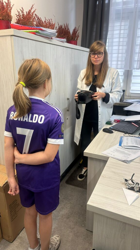
In every case, treatment is individually tailored to the patient’s needs and the severity of the disease. It is advisable to consult an ophthalmologist specializing in corneal diseases, who can help select the most appropriate treatment method.

Both surgical and non-surgical treatment options are available, and the choice depends on the advancement of keratoconus. In the early phases of the disease, non-invasive approaches are often preferred, while in more advanced cases surgical interventions may be necessary to stop disease progression.

The main treatment options include:

Glasses and contact lenses:
In mild cases of keratoconus, these may be sufficient to improve vision. Contact lenses—especially rigid gas-permeable lenses—can be effective in correcting corneal shape irregularities.

Corneal cross-linking (CXL):
A procedure designed to strengthen the corneal structure using ultraviolet light and vitamin B2 (riboflavin). CXL can slow down or stop the progression of keratoconus.

Corneal transplantation (keratoplasty):
In advanced cases where other treatments fail, a corneal transplant may be necessary. Different techniques are available, such as anterior lamellar keratoplasty or full-thickness corneal transplantation (penetrating keratoplasty).

Laser keratectomy:
In selected cases where keratoconus is not very advanced, laser keratectomy may be considered to help improve corneal shape and visual quality.

Other surgical procedures, such as intracorneal ring segments:
This procedure involves placing thin, arc-shaped rings within the cornea to improve its shape. The implanted rings exert pressure on the cornea, flattening the apex of the cone and making its shape closer to normal.

Do NOT rub your eyes!
Uncontrolled eye rubbing may occur as a symptom associated with keratoconus—it can be both a cause and a consequence of the disease, or a result of accompanying allergic conditions and atopy.

It is important to remember that early diagnosis and professional treatment of keratoconus significantly increase the chances of stopping disease progression and improving visual acuity. If you experience concerning symptoms suggestive of keratoconus in yourself or observe these in your teenage child, you should consult an ophthalmologist as soon as possible.